Contrast Agents
Authors
Brian Chung, MD
Assistant Professor of Anesthesiology
Honorio T. Benzon, MD
Professor of Anesthesiology
Department of Anesthesiology
Northwestern University Feinberg School of Medicine
Chicago, Illinois
Introduction
All body tissues attenuate the amount of radiation that reaches a radiographic detector to some degree. Contrast agents for conventional radiography are most commonly iodine-based. When placed in the body via any of a number of routes, these agents often provide greater attenuation than either the tissue or bone itself. This allows a differentiation between tissues that would otherwise have similar attenuation properties.
An ideal RCM agent has several properties including providing reasonable attenuation to delineate anatomic structures. The agent should be nontoxic to target tissues as well as non-target sites and should not be hazardous if injected into an unintended site. It should be able to linger long enough for radiographic study. On the other hand, the substance should have reasonably rapid clearance after sufficient images are obtained. The agent should also be cost-effective.
Indications
Radio-contrast media (RCM) facilitate several determinations including confirmation of correct needle or catheter placement. This is documented by either continuous fluoroscopic imaging during contrast administration or by comparison of pre- and post- injection images. Confirmation of location within correct structures often relies on a characteristic or typical distribution of RCM.
History
Contrast agents were first used at the Mayo Clinic in the 1920s. These original agents conferred radio-opacity via 3 iodine molecules attached to a fully substituted benzene ring.[1] On figure 1, the “R” group is generic as it can be any side chain.

Figure 1. Chemical Structure of a Contrast Agent
The first generation contrast agents (Figure 2) have been used in clinical practice since the 1950s.[1] They have a high osmolality secondary to unbound iodine. The osmolality can be up to 8 times the physiologic osmolality of blood. They are commonly called ionic agents due to the charge on the iodinated molecule. The RCM molecule is the anion and either sodium or meglumine is the cation.
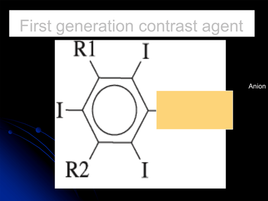
Figure 2. Chemical Structure of a First Generation Contrast Agent
The second generation contrast agents (Figure 3) have been used since 1986.[2] They were developed to avoid the potentially toxic cation. These are called nonionic agents. As the side chains are larger, they are more hydrophilic. Their osmolality is near-physiologic and they contain a minimal amount of free iodine. They have approximately the same viscosity and iodine concentration but less than half of the osmolality of the ionic agents. Of note, allergic reactions are less common with second generation agents compared to ionic agents. Currently, the clinical use of nonionic: ionic agents is 9:1.[3]
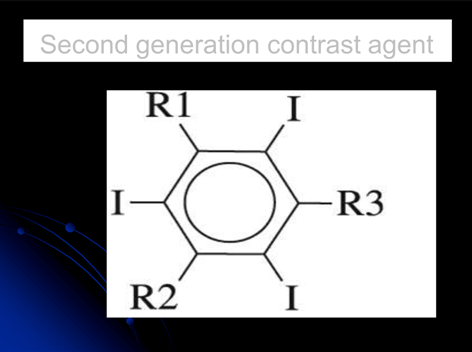
Figure 3. Chemical Structure of a Second Generation Contrast Agent
Complications
Adverse reactions are relatively uncommon with the second generation contrast agents. The overall incidence of adverse reaction is 1–2 percent for the low-osmolality agents compared to 5 percent for the high-osmolality agents. These reactions range from patient discomfort to hemodynamic instability. Physiologic toxicity is related to osmolality.[3] Adverse reactions typically occur within 15 minutes of exposure. These reactions may be chemotoxic, osmolar or allergic. Chemotoxic reactions include thyrotoxicosis, nephrotoxicity, and seizures.[3] Hyperosmolality can cause erythrocyte damage, endothelial damage, vasodilation of vasculature and cardiac depression. Many of these reactions are dose-dependent and the volumes given for needle guidance for spine procedures are typically relatively small.
Allergic Reactions
Allergic reactions may not be dose-related or infusion-rate-related. This reaction is actually anaphylactoid and is not IgE-mediated.[4] There is no evidence for an immunologic mechanism. RCM are one of the most commonly identifiable causes of anaphylactoid reactions in adults. Risk factors include prior allergic reaction to contrast, asthma, and atopy. It is very common for healthcare providers to equate shellfish allergy with iodine allergy. In actuality these are distinct reactions and represent two different pathophysiologies. As they are both relatively common, a false association has been perpetuated. Allergic reactions to shellfish are Ig-E mediated and the allergens are typically tropomyosin proteins.[5]
Clinical Features
The signs and symptoms of a contrast allergic reaction are principally caused by basophil and mast cell degranulation. These include bronchospasm, hypotension, urticaria, flushing, and angioedema. Allergic reactions are usually very mild and can be treated with minimal supportive care. However, reactions can also be life-threatening. Treatment remains supportive but they may require aggressive respiratory and hemodynamic resuscitation in addition to the use of antihistamines, corticosteroids and vasopressors.
For patients with a history of allergy to RCM, laboratory testing is not necessary. One should try to make a distinction between true allergic signs versus chemotoxic reactions. If a true allergy is suspected, one can defer the use of contrast agents realizing that the precise location or spread of injectate may be questionable. The patient can also be premedicated to avoid an anaphylactoid reaction. It is usually prudent to place an IV catheter, especially if the prior reaction was moderate to severe. Pretreatment protocols for patients with a history of RCM allergy are not universal, an example is shown in the table. These typically involve administration of prednisone, an H1 blocker, and an H2 blocker in a series of doses prior to the procedure 6. Patients must be monitored for 30-60 minutes post-procedure and emergency support should be immediately accessible.
Prevention
Pretreatment regimen for previous RCM allergic reactions is outline below.[7]
- 12 hours before procedure
- Prednisone, 20-50 mg p.o.
- Ranitidine, 50 mg p.o.
- Diphenhydramine, 25-50 mg p.o.
- hours before procedure
- Prednisone, 20-50 mg p.o.
- Ranitidine, 50 mg p.o.
- Diphenhydramine, 25-50 mg p.o.
- Immediate before procedure
- Diphenhydramine, 25 mg p.o.
Iodine-Based Contrast Agents
The most commonly used radio-contrast media agents include Omnipaque and Omnivue. They are preservative-free and water soluble. As part of their brand name there is usually a number which signifies the ionic concentration (in mg iodine/mL). There are formulations from each proprietary brand that are approved for intrathecal use and thus would be safe even if the administration were unintentional. Typically these agents are excreted in the urine with minimal metabolism and minimal protein-binding.
Non Iodine-Based Contrast Agents
Currently there are few alternatives to iodine-based RCM. If the patient has a moderate-to-severe iodine-contrast allergy, or if such patient is not premedicated on the day of the planned procedure, then one should consider the off-label use of a gadolinium chelate–based agent (e.g. Magnevist).[8] Small retrospective studies show that it may be a safe alternative. Of note these agents are FDA-approved for use with MRI and approved only for IV use. The image quality with these agents is typically lower than iodine-based contrast media. In addition, concentrations are greater than those used for MRI attenuation. Although rare, allergic reactions have been reported with gadolinium-based agents.[9] Since patients with history of allergic reactions are more likely to have other allergic manifestations, these patients need to be monitored for possible adverse reactions.
Summary
Overall, the use of RCM is safe for needle guidance for interventional pain medicine procedures. Most institutions are required to perform a Procedure Time-Out and this should be tailored to capture those patients with allergies to these agents so that appropriate adjustments may be made. Most reactions are not dose-related so it is prudent to use the smallest dose necessary that still allows for high-quality images. Vigilance should always be maintained to detect for adverse reactions to these agents.
References
- Kern MJ, King SB, Douglas JS. Cardiac catheterization, cardiac angiography, and coronary blood flow and pressure measurements. In: Fuster V, O’Rourke RA, Walsh RA, Poole-Wilson P, Eds. King SB, Roberts R, Nash IS, Prystowsky EN (eds.). Hurst’s The Heart, 11th Edn. New York:McGraw-Hill Companies, Inc, 2004, pp. 488.
- Costa N. Understanding contrast media. J Infusion Nursing 2004;27:302-12.
- Eng J. Contrast Studies. In Tintinalli JE, Kelen GD, Stapczynski JS, Ma OJ, Cline DM (eds.). Tintinalli’s Emergency Medicine: A Comprehensive Study Guide, 6th Edn. http://www.accessmedicine.com/content.aspx?aID=614899, accessed 07/09.
- Morcos SK. Acute serious and fatal reactions to contrast media: our current understanding. Brit J Rad 78(932):686-93, 2005 Aug.
- Sicherer SH. Risk of severe allergic reactions from the use of potassium iodide for radiation emergencies. J Allergy Clin Immunol 2004;114:1395-7
- Kelly JF, Patterson R, Lieberman P, et al. Radiographic contrast media studies in high-risk patients. J Allergy Clin Immunol 1978;62:181–184.
- Sitzman, BT, Chen Y, Rallo Clemans, Fishman SM, Benzon HT. Pharmacology for the interventional pain physician. In Benzon HT, Raja SN, Molloy RE, Liu SS, Fishman SM (eds). Essentials of pain medicine and regional anesthesia. 2nd Ed. Philadelphia: Elsevier Churchill-Livingston, 2005:166-180.
- Strunk HM, Schild H. Actual Clinical Use of Gadolinium-chelates for non-MRI applications. Eur Radiol 2004;14:1055-62.
- Safriel, Ang R, Ali M. Gadolinium Use in Spine Procedures for Patients with Allergy to Iodinated Contrast—Experience of 127 Procedures. AJNR 2006; 27:1194-7.
Leave a commentOrder by
Newest on top Oldest on top